Image Analysis | God is the creator. Doctors are the repairers! See how muscular skeletal ultrasound detects a little problem in the body that is only 0.03cm in size
November 28-2017

God is definitely a Virgo. He can use a 1.2% gene difference to distinguish humanity and orangutans, and can also use a little 0.03 cm problem to put people in unbearable pain. 


How small is 0.03 cm? It is not visible to the naked eye. There is no way to sense it by touch. However, to body repairers ---- clinical physicians, a piece of ultrasound equipment equipped with a high frequency probe can help them discern what’s truly wrong. 


Muscular Skeletal Ultrasound


A New Weapon for Rehabilitation Doctors 

Muscular skeletal ultrasound has been a burgeoning field of ultrasound applications in recent years, and is regarded as one of the development directions with the most potential. Muscular skeletal ultrasound can assist doctors in achieving dynamic observation of tendon and intramuscular activity, and is also able to conduct interventional therapy through the guidance of color Doppler ultrasound, and avoid blind insertion failures or the triggering of complications by injecting the medicine into the outside of the tendon sheath and the inside of the tendon. It greatly improves clinical diagnostic efficiency and precision. 
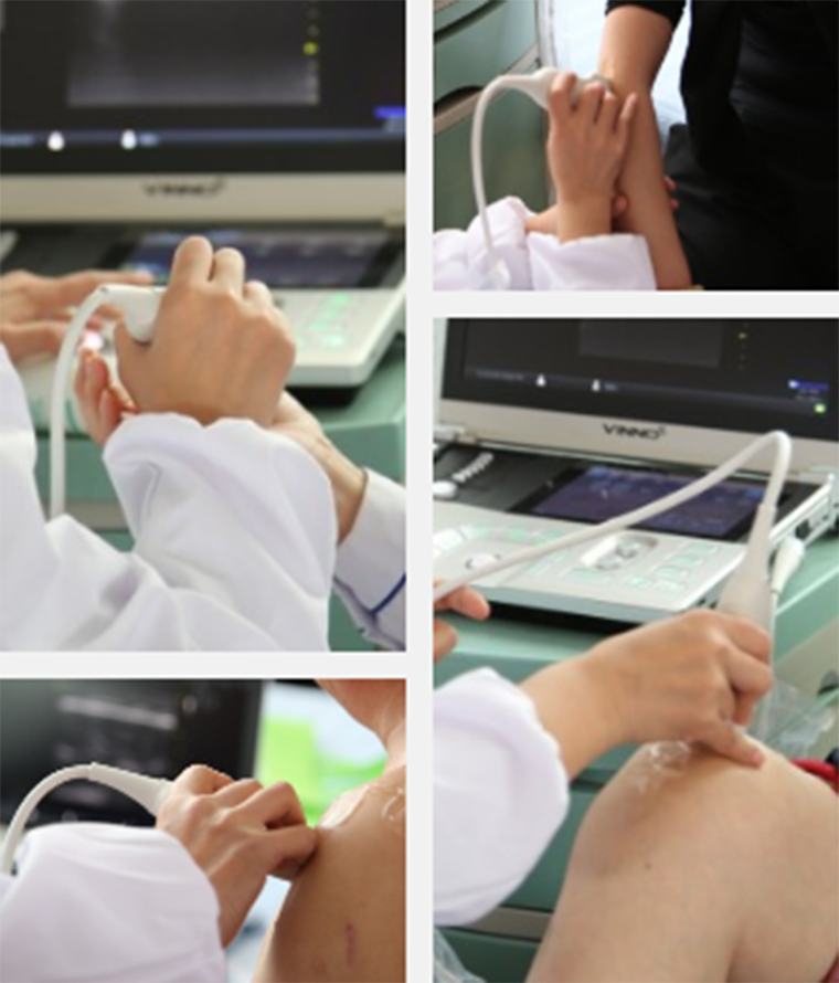
Muscle, tendons, ligaments joints, and peripheral nerves can all be precisely diagnosed. 

Based on muscular skeletal ultrasound’s advantages of convenience and precision Shandong province’s city of Weifang organized a physical rehabilitation summit forum and charity clinic event. This event was sponsored by the local district government, the district Hygiene and Family Planning Bureau, the district China Merchants Group, the district Athletics Bureau, and the Qingdao Century Jiechuang Medical Technology Company, and was organized by the district People’s Hospital. As a high-end national color Doppler ultrasound brand VINNO provided equipment support. The groups of people at the event had an enthusiastic reaction. Even the doctors from the local hospitals all came to get a checkup. 

And so the problems came up. What are the primary uses for muscular skeletal ultrasound? What types of diseases can you consider after seeing what kinds of images? Please view the following real life examples↓


Ultrasound Image Collection


Muscle, Tendon, Ligament, Joint Capsule, etc

Case 1:

Image displays no connection with the tendon. The consideration is that there is a solid-cystic tumor at the back of the left hand

Case 2:
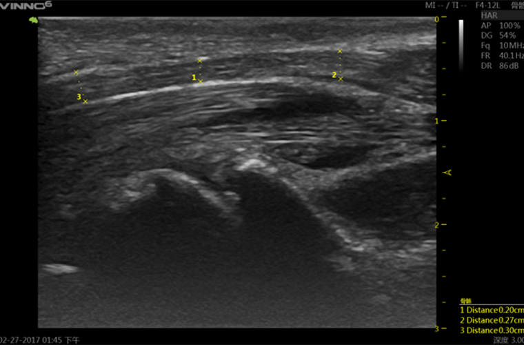
Image displays pressure on the median nerve. The diagnosis is carpal tunnel syndrome.

Case 3:

Image displays that the burca mucosa behind the heel bone is accumulating fluid. The diagnosis is Achilles tendon enthesopathy. 

Case 4:

Image displays uratoma of the toe joint. The diagnosis is gout. 

Case 5:
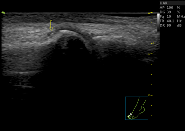
The first metatarsophalangeal joint. The arrow points to a urate crystal on the first metatarsophalangeal joint. The diagnosis is gout. 

Case 6: 

Image displays a ganglion cyst on the back of the foot. 

Case 7:

Image displays calcification adhered to the triceps brachii muscle tendon.

Case 8:
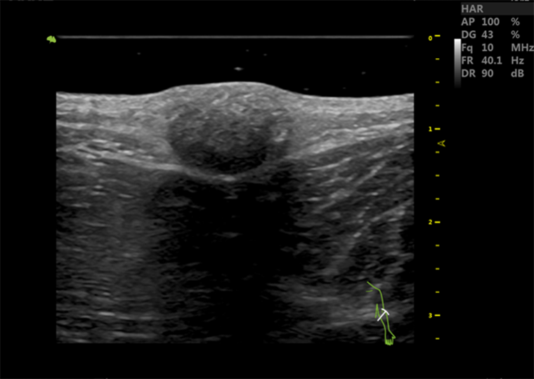
Image displays a solid mass. The consideration is that it is possibly pilomatricoma.

Case 9:

Image displays a large amount of fluid accumulation at the knee joint cavity. The consideration is that it was caused by Ankylosing Spondylitis (AS). 

Case 10:

 Image displays fluid accumulation at the knee joint cavity. The diagnosis is synovitis. 


Of course, using muscular skeletal ultrasound to discover a problem is only the first step. The precise physical diagnosis system for muscular skeletal pain that this charity clinic used also included a high energy laser and an extracorporeal shock wave (ESW) therapeutic regime, easing the muscular skeletal pain of patients on site. 


The World’s Most Cutting Edge and Most Effective


Precise Physical Diagnosis System for Muscular Skeletal Pain


The precise physical diagnosis system for muscular skeletal pain is currently the most effective solution for muscular skeletal pain. The diagnostic procedure is split up into three parts:

1 Muscular skeletal ultrasound precisely diagnoses the afflicted part of the body and the causes of the affliction.

2 The high energy laser eases pain, diminishes inflammation, and eliminates edema.

3 The extracorporeal shock wave repairs, relieves, and treats pain. 

The charity clinic specially invited Mark Callanen, an American physical therapist, a PhD in clinical physical therapy, and a clinical orthopedic specialist, to conduct treatment for the patients. 


This solution that requires no surgery, offers a precise diagnosis, and which has quick results is already in service at institutions like the State General Administration of Sports, the Beijing 301 Hospital, and the Peking University of Health Science Center’s Number Three Hospital. The charity clinic has brought this solution to the people in the community. After treatment most of the patients expressed that their pain had been relieved. 
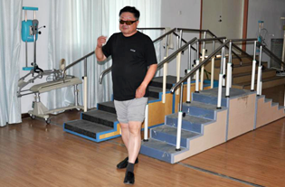
 

A patient’s knee joint cavity pain was eased. Climbing up the stairs was easy. 


Benefiting humanity with the most advanced medical technology, VINNO is willing to resolve the tiny ailments of the human body together with doctors. God is the creator. Doctors are the repairers. A high-end national color Doppler ultrasound brand is the top choice of rehabilitation physicians domestically and internationally! 


Return list
Image Analysis | God is the creator. Doctors are the repairers! See how muscular skeletal ultrasound detects a little problem in the body that is only 0.03cm in size
November 28-2017

God is definitely a Virgo. He can use a 1.2% gene difference to distinguish humanity and orangutans, and can also use a little 0.03 cm problem to put people in unbearable pain. 


How small is 0.03 cm? It is not visible to the naked eye. There is no way to sense it by touch. However, to body repairers ---- clinical physicians, a piece of ultrasound equipment equipped with a high frequency probe can help them discern what’s truly wrong. 


Muscular Skeletal Ultrasound


A New Weapon for Rehabilitation Doctors 

Muscular skeletal ultrasound has been a burgeoning field of ultrasound applications in recent years, and is regarded as one of the development directions with the most potential. Muscular skeletal ultrasound can assist doctors in achieving dynamic observation of tendon and intramuscular activity, and is also able to conduct interventional therapy through the guidance of color Doppler ultrasound, and avoid blind insertion failures or the triggering of complications by injecting the medicine into the outside of the tendon sheath and the inside of the tendon. It greatly improves clinical diagnostic efficiency and precision. 

Muscle, tendons, ligaments joints, and peripheral nerves can all be precisely diagnosed. 

Based on muscular skeletal ultrasound’s advantages of convenience and precision Shandong province’s city of Weifang organized a physical rehabilitation summit forum and charity clinic event. This event was sponsored by the local district government, the district Hygiene and Family Planning Bureau, the district China Merchants Group, the district Athletics Bureau, and the Qingdao Century Jiechuang Medical Technology Company, and was organized by the district People’s Hospital. As a high-end national color Doppler ultrasound brand VINNO provided equipment support. The groups of people at the event had an enthusiastic reaction. Even the doctors from the local hospitals all came to get a checkup. 

And so the problems came up. What are the primary uses for muscular skeletal ultrasound? What types of diseases can you consider after seeing what kinds of images? Please view the following real life examples↓


Ultrasound Image Collection


Muscle, Tendon, Ligament, Joint Capsule, etc

Case 1:

Image displays no connection with the tendon. The consideration is that there is a solid-cystic tumor at the back of the left hand

Case 2:

Image displays pressure on the median nerve. The diagnosis is carpal tunnel syndrome.

Case 3:

Image displays that the burca mucosa behind the heel bone is accumulating fluid. The diagnosis is Achilles tendon enthesopathy. 

Case 4:

Image displays uratoma of the toe joint. The diagnosis is gout. 

Case 5:

The first metatarsophalangeal joint. The arrow points to a urate crystal on the first metatarsophalangeal joint. The diagnosis is gout. 

Case 6: 

Image displays a ganglion cyst on the back of the foot. 

Case 7:

Image displays calcification adhered to the triceps brachii muscle tendon.

Case 8:

Image displays a solid mass. The consideration is that it is possibly pilomatricoma.

Case 9:

Image displays a large amount of fluid accumulation at the knee joint cavity. The consideration is that it was caused by Ankylosing Spondylitis (AS). 

Case 10:

 Image displays fluid accumulation at the knee joint cavity. The diagnosis is synovitis. 


Of course, using muscular skeletal ultrasound to discover a problem is only the first step. The precise physical diagnosis system for muscular skeletal pain that this charity clinic used also included a high energy laser and an extracorporeal shock wave (ESW) therapeutic regime, easing the muscular skeletal pain of patients on site. 


The World’s Most Cutting Edge and Most Effective


Precise Physical Diagnosis System for Muscular Skeletal Pain


The precise physical diagnosis system for muscular skeletal pain is currently the most effective solution for muscular skeletal pain. The diagnostic procedure is split up into three parts:

1 Muscular skeletal ultrasound precisely diagnoses the afflicted part of the body and the causes of the affliction.

2 The high energy laser eases pain, diminishes inflammation, and eliminates edema.

3 The extracorporeal shock wave repairs, relieves, and treats pain. 

The charity clinic specially invited Mark Callanen, an American physical therapist, a PhD in clinical physical therapy, and a clinical orthopedic specialist, to conduct treatment for the patients. 


This solution that requires no surgery, offers a precise diagnosis, and which has quick results is already in service at institutions like the State General Administration of Sports, the Beijing 301 Hospital, and the Peking University of Health Science Center’s Number Three Hospital. The charity clinic has brought this solution to the people in the community. After treatment most of the patients expressed that their pain had been relieved. 


A patient’s knee joint cavity pain was eased. Climbing up the stairs was easy. 


Benefiting humanity with the most advanced medical technology, VINNO is willing to resolve the tiny ailments of the human body together with doctors. God is the creator. Doctors are the repairers. A high-end national color Doppler ultrasound brand is the top choice of rehabilitation physicians domestically and internationally! 


Return list